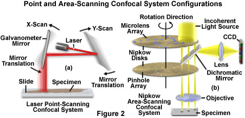
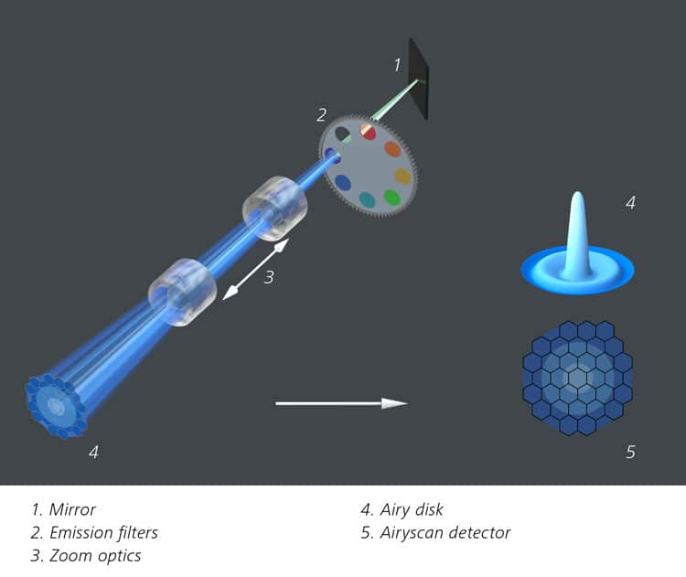
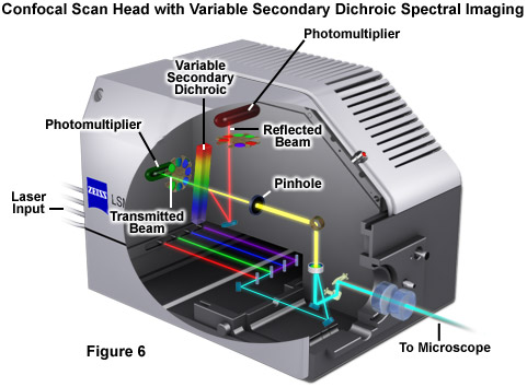
Confocal microscopy most frequently confocal laser scanning microscopy clsm or laser confocal scanning microscopy lcsm is an optical imaging technique for increasing optical resolution and contrast of a micrograph by means of using a spatial pinhole to block out of focus light in image formation.
Benefits of laser scanning microscopy.
In this interview olympus dr.
The benefits of delivering higher efficiency imaging at lower laser powers include less photobleaching phototoxicity and is less expensive than confocal laser scanning microscopes.
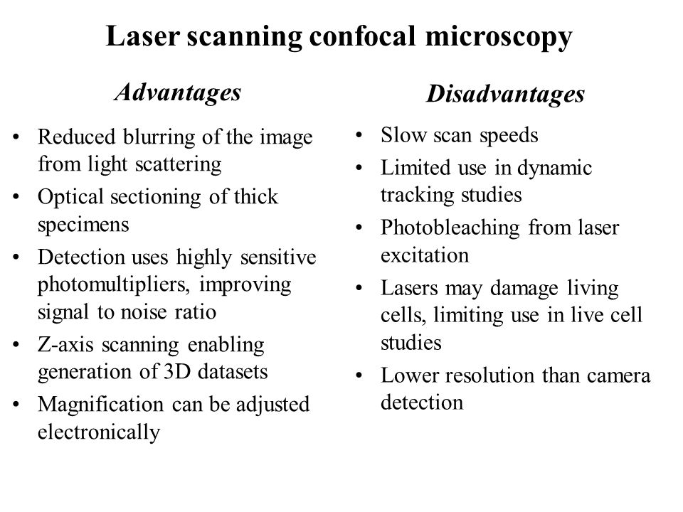
Advantages of confocal laser scanning microscopy industrial applications of confocal microscopy thin film profiling.
Confocal microscopy is a powerful tool that can be used to create 3d images of the structures within living cells and to examine the dynamics of cellular processes 1 for high speed imaging of fluorescent molecules and structures within living cells microscopes must provide rapid field of view scanning that eliminates any out of focus.
When imaged with a laser scanning confocal microscope figure 1 d the.
Benefits of confocal microscopy.
Laser scanning confocal types can cut exceptionally clean thin optical sections of about 0 5 to 1 5 µm out of thick samples measuring more than 50 µm using.
Clsm combines high resolution optical imaging with depth selectivity which allows us to do optical sectioning.
Additional benefits of laser confocal microscopy include fast image acquisition and high resolution microscope images over a wide area which can help save valuable testing and production time.
See what she has to say about precision ease of use and other capabilities that make confocal microscopes invaluable inspection tools.
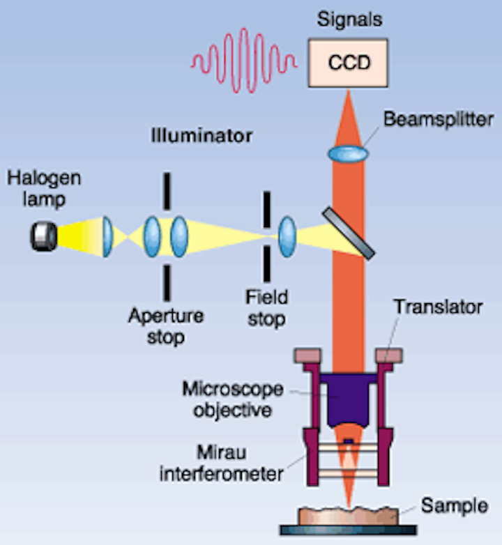
The thickness of the coating can be determined by observing the 2 peaks in the axial intensity variation.
The primary advantage of laser scanning confocal microscopy is the ability to serially produce thin 0 5 to 1 5 micrometer optical sections through fluorescent specimens that have a thickness ranging up to 50 micrometers or more.
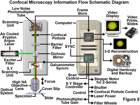
Laser confocal microscope focus detection system.
Benefits of confocal microscopy for live cell imaging.
When investigating multilayer structures the true surface of a substrate can be observed through a surface coating.
Capturing multiple two dimensional images at different depths in a sample enables the.
A thick section of fluorescently stained human medulla in widefield fluorescence exhibits a large amount of glare from fluorescent structures above and below the focal plane figure 1 a.
Laser scanning confocal fluorescence microscopy.
Fluorescent microscopy not only makes our images look good it also allows us to gain a better understanding of cells structures and tissue.
With confocal laser scanning microscopy clsm we can find out even more.
This means that we can view visual sections of tiny structures that.




.png)






.jpg)
